

Quick Summary
- Lumbar spinal stenosis is a narrowing of the spinal canal in the lower back that puts pressure on the nerves traveling into your legs and buttocks.
- The hallmark symptom is leg pain, heaviness, or tingling that comes on during standing or walking and eases when you sit down or lean forward.
- A scan showing canal narrowing does not automatically mean symptoms will be severe or that surgery is needed; diagnosis requires matching imaging to your clinical picture.
- Most people begin with physical therapy, exercise, activity pacing, and medication, and many improve meaningfully without an operation.
- Surgery is reserved for people whose walking or daily independence is substantially limited after conservative treatment, or who have worsening nerve problems in their legs.
For many adults over 60, everyday activities like grocery shopping or a walk around the block become genuinely difficult because of pressure building on the nerves in the lower back. Lumbar spinal stenosis is a frequent reason for referral to a spine specialist, and its effects can range from persistent leg heaviness to walking that forces a rest stop every few minutes. Most people improve with the right care, and surgery is only one of several options. This guide covers what lumbar spinal stenosis is, how it’s recognized and confirmed, what conservative treatment involves, and when an operation becomes a reasonable consideration.
What Is Lumbar Spinal Stenosis?
Lumbar spinal stenosis is a narrowing of the spinal canal or the smaller openings where nerve roots exit the spine in the lower back. That narrowing squeezes the spinal nerves and causes the pain, numbness, or weakness most patients feel in the buttocks and legs. The condition most commonly develops from gradual, age-related changes in the spine rather than from a single injury or event.
Three specific areas of the lumbar spine can narrow. The central canal runs down the middle of the vertebral column and contains the spinal cord and the cauda equina, which is the bundle of nerve roots below the spinal cord. The lateral recess is a smaller pocket just before each nerve root turns outward toward the leg, and the foramen is the opening through which each nerve root exits the spine on its way to the lower limb. Any combination of these three can be involved at the same time, at one or more spinal levels.
Age-related wear is the leading cause. As the disc between two vertebrae loses height, the facet joints at the back of the spine take on more load and gradually enlarge. The ligamentum flavum, a tough band of tissue running along the back of the canal, thickens over years of use. Together, these changes progressively close off the space nerves need. The NINDS spinal stenosis resource identifies these degenerative changes as the most common cause of lumbar spinal stenosis in adults.
Less often, it follows scoliosis, a prior spinal injury, or previous spine surgery. The AAOS patient education page on lumbar spinal stenosis notes that imaging can show canal narrowing in people with no symptoms at all, which is why treatment decisions should rest on both the scan findings and the patient’s overall clinical picture.
How the Condition Feels: Recognizing the Patterns
The defining symptom pattern of lumbar spinal stenosis is neurogenic claudication: pain, heaviness, tingling, numbness, or weakness that builds in the buttocks and legs during standing or walking and reliably eases when you sit down or lean forward. Recognizing this pattern is what separates lumbar spinal stenosis from most other causes of leg discomfort.
The relief from leaning forward has a mechanical explanation: bending the lower spine forward slightly increases the space inside the canal, taking pressure off the compressed nerves. That’s why many patients find walking uphill easier than walking on flat ground, and why cycling, which keeps the lumbar spine in a gently flexed position, is often better tolerated than brisk walking on a level surface.
- Aching, burning, or cramping in one or both legs during walking, often building after a certain distance and forcing a rest stop.
- Numbness or tingling that radiates from the lower back into the buttocks, thighs, or calves.
- Leg heaviness or weakness that accumulates with standing time and progressively shortens how far you can walk.
- Low-back pain that may be less disabling than the leg symptoms for many patients, particularly as the condition advances.
Neurogenic claudication needs to be distinguished from vascular claudication, which is leg pain caused by poor blood flow rather than nerve compression. Vascular claudication typically doesn’t ease with leaning forward and doesn’t worsen with simply standing still. A clinician can usually tell the two apart with a targeted history and a physical examination. Peripheral neuropathy from diabetes can produce similar tingling and numbness, but it follows a different symptom pattern and doesn’t respond to postural changes the same way.
How Doctors Diagnose Spinal Stenosis
A reliable diagnosis of lumbar spinal stenosis requires a detailed account of how and when your symptoms appear, a neurological assessment covering strength, reflexes, sensation, and gait, and imaging that confirms the narrowing corresponds to the reported complaints. A scan showing canal narrowing without matching symptoms is not, by itself, a diagnosis that requires treatment.
During the physical examination, a clinician checks muscle strength, tendon reflexes, and sensation in the legs and feet. They watch your gait and may ask you to walk a measured distance to see whether and when leg symptoms appear. The NIAMS back pain resource emphasizes that a careful evaluation of symptoms and physical findings should guide treatment planning far more reliably than imaging alone.
MRI of the lumbar spine is the standard imaging study for spinal stenosis. It shows the spinal canal, nerve roots, intervertebral discs, and ligaments in detail without radiation. Standing X-rays reveal overall spinal alignment, disc height, and whether one vertebra has slipped forward on the one below, a condition called spondylolisthesis. When MRI isn’t suitable, a CT scan provides detailed views of the bony canal structure. Electromyography (EMG) and nerve-conduction studies may be added when a clinician wants to rule out peripheral neuropathy or another nerve disorder that could independently explain the leg symptoms.
A clinical reference chapter in StatPearls, available at NCBI Bookshelf (NBK531493), notes that visible canal narrowing on MRI is common among adults over 60 even without any symptoms, which underscores why imaging findings alone cannot determine whether or how to treat.
| Diagnostic Tool | What It Shows | When It Is Typically Used |
|---|---|---|
| MRI | Soft tissue, nerves, discs, ligaments, canal width | First-choice imaging for most patients |
| Standing X-ray | Alignment, disc height, spondylolisthesis | Initial evaluation and surgical planning |
| CT scan | Detailed bone structure and canal shape | When MRI is unavailable or contraindicated |
| EMG and nerve-conduction study | Nerve function, peripheral neuropathy screening | When another nerve disorder is suspected |
| Physical examination | Strength, reflexes, sensation, gait, walking tolerance | Every evaluation; guides imaging interpretation |
Nonsurgical Treatment: Physical Therapy, Medication, and Injections
Most people with lumbar spinal stenosis begin with conservative care. Physical therapy, activity modification, appropriate medication, and sometimes epidural steroid injections can meaningfully reduce pain and restore walking ability without an operation. None of this reverses the structural narrowing, but these measures can improve strength, balance, endurance, and daily function for a large share of patients.
Physical therapy is the backbone of nonsurgical management. A physical therapist builds an individualized program that typically includes exercises to strengthen the deep abdominal and back muscles, gentle lumbar flexion movements that open space inside the canal, and balance and gait training to cut fall risk. Aquatic therapy is often well tolerated because water reduces the load through the spine while still allowing effective exercise. Consistent participation in a supervised program, rather than sporadic effort on good days only, generally produces the best results.
Activity modification means pacing your day to avoid prolonged standing or walking without planned rest breaks. A walking stick, rollator frame, or shopping cart provides a forward lean that many patients find noticeably reduces leg symptoms during movement. Choosing routes with benches, breaking tasks into shorter intervals, and resting before pain becomes severe can extend your comfortable daily range considerably.
Medications used for symptom relief in lumbar spinal stenosis include nonsteroidal anti-inflammatory drugs (NSAIDs) for pain and inflammation, and neuropathic agents when burning or electric-shock sensations are prominent. Any medication plan should be reviewed carefully with a clinician or pharmacist, especially in older adults who may be taking several other drugs or who have kidney, stomach, or cardiovascular concerns that affect which medications are safe to use.
Epidural steroid injections place anti-inflammatory medication directly around the compressed nerve roots in the lumbar spine. The Mayo Clinic treatment overview notes that injections may provide short-term symptom relief for selected patients, though they don’t permanently widen the spinal canal and their benefit tends to diminish with repeated use over time.
Tip: Keeping a brief diary of your pain level and walking distance before each clinic visit helps your doctor track whether conservative treatment is working and decide when to adjust the plan.
When Is Surgery Recommended for Spinal Stenosis?
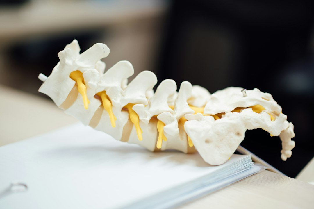
Surgery for lumbar spinal stenosis is generally considered when symptoms substantially limit walking or daily activities, when consistent nonsurgical treatment over several months has not provided adequate relief, or when progressive neurologic problems are developing in the legs. It’s not the right first step for most patients, but it becomes a reasonable option when those specific conditions are met.
Decompression surgery removes the bone spurs, thickened ligament, or other tissue pressing on the nerves. A laminectomy, the most common decompression procedure, removes part of the vertebral arch at the back of the spine to widen the canal. The goal is to relieve nerve pressure. As the AAOS patient education page notes, leg pain and walking limitation tend to improve more reliably after decompression than nonspecific low-back pain does, and patients should understand this difference before agreeing to surgery.
Spinal fusion is a separate procedure that may be added to decompression in some cases. It joins two or more vertebrae together permanently and is generally considered when instability, spondylolisthesis, deformity, or another specific finding makes decompression alone unsuitable. Fusion carries its own recovery timeline and considerations, and it’s not a routine addition to every decompression procedure.
For older adults with other health conditions such as heart or lung disease, the surgical decision requires careful shared discussion with a spine surgeon. Risks must be weighed against potential benefits on an individual basis. Symptom severity, the impact on daily independence, personal goals, and overall health all figure into that conversation alongside the structural findings on imaging.
Tip: Ask your surgeon specifically which of your symptoms, such as leg pain, walking limitation, or back ache, are most likely to improve after the planned procedure. A clear answer before surgery helps set realistic expectations for recovery.
Recovery, Outlook, and Protecting Long-Term Mobility
Recovery after lumbar spinal stenosis treatment varies depending on whether surgery or conservative care was pursued, your overall health, and how consistently you engage with rehabilitation. Setting realistic expectations about which symptoms are likely to improve, and over what timeframe, makes a real difference in how well you work through the treatment plan.
After decompression surgery, leg pain and the ability to walk further are often among the first things to improve. Back pain may take longer to settle or may not fully resolve, because decompression targets nerve compression rather than the underlying degenerative changes in the discs and facet joints. Supervised rehabilitation typically begins once the surgical team confirms that healing is on track, and progresses at a pace your surgeon and physiotherapist set together.
When fusion is added alongside decompression, the recovery period is longer. The spine needs adequate time to heal and form a solid bone bridge between the fused segments. Physical therapy progresses gradually and is important for rebuilding strength, confidence with movement, and walking endurance.
For patients who manage without surgery, consistent physical therapy and activity modification often produce real improvement in pain, walking distance, and daily function over time. The structural narrowing doesn’t reverse, but strengthening the muscles that support the lumbar spine reduces the load on compressed structures and helps many people maintain function for longer.
Fall risk may be higher in older adults with lumbar spinal stenosis, as leg weakness and reduced balance can increase the chance of a fall, which can lead to a separate and serious injury. Balance exercises from a physiotherapist, appropriate footwear with good grip, and a simple home safety review are practical steps worth raising at your next appointment.
When to See a Doctor
See a doctor soon if:
- Your walking distance is shortening noticeably over weeks and limiting your daily independence.
- Leg pain, tingling, or weakness is spreading or intensifying despite several weeks of home management and activity adjustment.
- You are relying on pain medication most days without lasting relief.
- You have had a recent fall or feel unsteady on your feet due to leg weakness.
Seek emergency care immediately if:
- You develop new difficulty controlling your bladder or bowel.
- You notice numbness or tingling in the area between your legs (the saddle area).
- You experience sudden, rapidly worsening weakness in one or both legs.
- You have severe and quickly progressing neurologic symptoms in the lower limbs.
These urgent signs can indicate cauda equina syndrome, a rare but serious condition that requires immediate medical assessment and must not wait for a scheduled appointment.
Key Takeaways
- Lumbar spinal stenosis is age-related narrowing of the lower-back spinal canal that presses on nerves, most often from a combination of enlarged facet joints, disc-height loss, and thickened ligaments rather than a single cause.
- The hallmark symptom pattern is leg heaviness, tingling, or cramping that worsens with standing and walking and reliably improves by sitting or bending forward, reflecting increased canal space in a flexed posture.
- Diagnosis requires matching imaging findings to the patient’s symptoms and physical examination; canal narrowing visible on an MRI alone is not a sufficient basis for deciding on treatment.
- Conservative management, including supervised physical therapy, flexion-based exercises, activity pacing, and medication review, is the appropriate starting point for most patients and helps many regain meaningful function over weeks to months of consistent effort.
- Decompression surgery most reliably reduces leg pain and extends walking tolerance; nonspecific low-back pain improves less predictably; fusion is reserved for specific indications such as spondylolisthesis or spinal instability.
- New bowel or bladder problems, saddle numbness, or rapidly worsening leg weakness are emergency warning signs that require immediate assessment, not a routine clinic appointment.
Conclusion
Lumbar spinal stenosis is a common and manageable condition, particularly when caught early and treated in a thoughtful, stepwise way. Understanding the link between your symptoms and the underlying canal narrowing puts you in a stronger position when talking with your doctor about the best path forward.
For most people, treatment begins with physical therapy, targeted exercises, and adjustments to daily activity. These measures don’t undo the structural narrowing, but they can substantially improve comfort, walking distance, and quality of life for a large share of patients. When conservative care isn’t enough, decompression surgery is a well-established option for appropriately selected patients, with its clearest and most consistent benefits coming in leg pain relief and extended walking tolerance.
Treatment decisions should be based on how your symptoms affect your daily life, not only on what a scan shows. Staying active within your limits, attending scheduled physiotherapy sessions, and keeping your clinical team informed of any changes all contribute to better long-term outcomes. If your symptoms are shifting or worsening, refer back to the warning signs described in this guide and contact your doctor promptly.
Frequently Asked Questions
What is lumbar spinal stenosis and what causes it?
Lumbar spinal stenosis is a narrowing of the spinal canal or nerve-root openings in the lower back that compresses spinal nerves. The most common cause is age-related wear, including facet-joint enlargement, disc-height loss, and thickening of the spinal ligaments. Less often, scoliosis, prior injury, or previous spine surgery plays a role.
What symptoms in the legs point toward spinal stenosis?
The most recognizable pattern is neurogenic claudication: aching, heaviness, tingling, or weakness in the buttocks and legs that builds during standing or walking and reliably eases with sitting or leaning forward. This postural response distinguishes spinal stenosis from most other causes of leg pain.
Can lumbar spinal stenosis improve without surgery?
Many people manage well with nonsurgical care, including physical therapy, targeted exercises, activity pacing, and appropriate medication. Epidural steroid injections may offer short-term relief for some patients. Conservative treatment cannot reverse structural narrowing, but it commonly reduces pain and restores meaningful daily function.
When should someone consider surgery for lumbar spinal stenosis?
Surgery is generally considered when symptoms substantially limit walking or daily activities, when consistent nonsurgical treatment has not provided adequate relief, or when progressive neurologic deficits are developing. Decompression surgery most reliably addresses leg pain and walking limitation rather than nonspecific back pain.
What is recovery like after lumbar spinal stenosis surgery?
Recovery depends on whether decompression alone or decompression plus fusion was performed. Decompression-only procedures typically allow an earlier return to light activity, while fusion requires a longer healing period. Leg pain and walking distance often improve before back pain does, and supervised physical rehabilitation is an important part of recovery.
This article is for informational purposes only and does not replace professional medical advice. Always consult your doctor before starting any treatment.